


One of the most common diseases today isosteochondrosis of the lumbar spine. Both men and women alike often turn to doctors with this disease.
At least once in our life, each of us has experienced pain in the lower back, sacrum or lower leg. We often unconsciously call this type of pain "a pinched nerve in the lower back. "
Causes of lumbar osteochondrosis
According to research by doctors and scientists, the main cause of osteochondrosis of the lumbar spine is upright posture. However, not everyone has osteochondrosis. Provocative factors that accelerate the development of the disease are: metabolic disorders, physical inactivity, injury, excess weight, improper weight lifting.
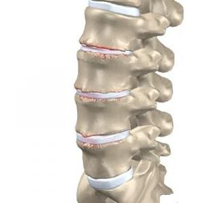
The source of pain in osteochondrosis is a pinched nerve root, which occurs after the protrusion of the intervertebral disc and the narrowing of the intervertebral gap. This deviation is formed when nutrition deteriorates, the natural process of oxygen and lymph exchange in the tissue of the intervertebral disc is disturbed. As a result, the shock absorption capacity of the intervertebral disc decreases, and the nucleus pulposus of the disc gradually decreases and dries up.
From an anatomical point of view, lumbar osteochondrosis is a process of transformation of cartilage into bone, resulting in excessive pressure on the nerve roots extending from the spinal cord. These changes cause pain. Excessive growth of bone tissue occurs as a result of nutritional deterioration of the intervertebral disc, loss of fluid and disturbances in structure and function.
When the protrusion of the intervertebral disc during the development of osteochondrosis of the lumbar spine becomes more serious, it gives rise to the development of lumbar protrusion and herniation of the lumbar disc due to the rupture of the fibrous ring.
The pain syndrome is formed due to pinching of the spinal nerves in lumbar osteochondrosis and is called lumboischialgia. This symptom is accompanied by numbness in the lower part of the leg. Depending on the nature and location of the pain, this disease can be divided into sciatica and lumbago. Inflammation of the nerve due to its pinching is called radiculitis. The method of treating radiculitis with painkillers is, in fact, only the elimination of symptoms, and is ineffective, because such treatment does not affect the real cause of the disease, which is the degenerative process in the intervertebral disc. To eliminate pain and take preventive measures to prevent complications, you need to undergo a comprehensive course of treatment to activate the recovery process in the disc tissue, normalize the height and physiological parameters of the intervertebral disc.
Symptoms of osteochondrosis of the lumbar spine
One of the signs of osteochondrosis is compression of the spinal nerve root by the nucleus pulposus protruding from the intervertebral disc. This compression occurs in the epidural space, which is a kind of container for the spinal roots. Osteochondrosis of the lumbar spine is indicated by the following symptoms corresponding to a compressed root:
- L1 and L2 - loss of sensitivity in the "rider pants" area, i. e. in the groin and inner thigh area. Pain can occur in both legs at once if lumbar osteochondrosis is complicated by the development of a hernia.
- L5 – shooting pain, decreased sensitivity in the lower back and thumb sensitivity, as well as decreased ability to flex the fingers.
- S1 – shooting pain, decreased sensitivity of the lower leg and outer thigh, pain in the foot from the big toe to the fourth toe. Often when this root is damaged, the Achilles and plantar reflexes are lost.
- Damage to the Deproge-Gotteron artery - in the chronic course of osteochondrosis, paralysis of the lower legs and back may occur, and sensitivity may be lost in the anogenital area.
- Simultaneous damage to the roots of L5, S and the Deproge-Gotteron artery causes the "paralyzed sciatica" syndrome, loss of pelvic and motor function.
Osteochondrosis of the lumbar spine can cause protrusion and herniation due to a significant load on this part of the spine. These complications develop quickly, so it is very important to treat them in time. Do not delay your visit to a vertebrologist, undergo a full examination, and seek qualified help at the first symptoms of lumbar osteochondrosis.
Complications of osteochondrosis of the lumbar spine
Compression-vascular ischemia can be considered a complication of lumbar osteochondrosis. This pathology develops due to impaired blood supply to the spinal cord, reduction of intervertebral openings for channels and arteries, as well as narrowing of the peripheral structures of the vertebrae. The reason for the development of this pathology is flattening of the disc, excessive mobility of the spine, weak ligaments, formation of osteophytes and neoarthrosis. Injury and permanent pressure on the pinched vessel or artery occurs due to any movement of the spine affecting the damaged area. In addition, reflex narrowing of the vessels that pass through the opening of the clamped channel may develop. This effect is called "narrow bed".
Vertebrologists identified another serious complication of osteochondrosis of the lumbar spine. Compressive myelopathy is a spinal cord disorder that occurs due to narrowing of the spinal canal. Depending on the location, the symptoms and severity of the pathology may vary. Often, the course of the disease is characterized by episodic - after an attack there is a period of remission.
Lumbar osteochondrosis can be complicated by TXII-L1 herniated disc, which exerts pressure and causes damage to the S1-S2, L2-L-4 segments of the spinal cord. Patients who experience this complication feel pain in the lower back, lower legs, back of the thighs, as well as weakness in the legs. The gluteal and calf muscles gradually become hypotonic and hypotrophic, the Achilles and plantar reflexes fall, and leg paresis develops. The back and outer surface of the foot and lower leg are characterized by reduced sensitivity or its complete absence.
If LI-II disc herniation develops, where excessive pressure on the S3 coccygeal segment increases, osteochondrosis of the lumbar spine has the following symptoms: disorders of the pelvic organs, fecal and urinary incontinence, constipation, loss or decreased sensitivity of the anogenital area, development of bed sores, reflexesanal prolapse.
"Cauda equina syndrome" develops when the nerve root is compressed from the first lumbar vertebra downwards. Experienced vertebrologists note that this syndrome most often develops in patients with congenital narrowing of the spinal canal. "cauda equina" is a group of nerve roots containing the terminal roots of the spinal cord from the first segment onwards. The name arises from its resemblance to a horse's tail. The diagnosis of this syndrome largely depends on the presence of unusual pain that is radicular in nature. This feature distinguishes cauda equina syndrome from other complications where there is no acute pain.
Cauda equina syndrome is characterized by severe pain in the sacrum and lower back, which radiates to the buttocks, anogenital area, and the back surface of the thighs. In some cases, paresis, peripheral paralysis, and sensory disturbances occur. Severe cases are characterized by paralysis of the back and both legs. A characteristic feature of complications can be considered asymmetry of sensory and motor disturbances.
Lumbar osteochondrosis can cause the development of myelogenous "intermittent claudication" syndrome, in which there is weakness in the legs when walking, a frequent urge to urinate, and numbness in the lower part of the body. The cause of this discomfort is poor blood supply to the lower part of the spinal cord. After resting, this sensation disappears.
With cauda equina root ischemia, caudogenic "intermittent claudication" occurs - goosebumps and tingling sensations when walking. Over time, these symptoms can rise higher, reaching the perineum, genitals and groin. Weakness in the legs disappears after a short rest.
Compression of the Adamkiewicz artery can occur due to unsuccessful sudden movements, heavy lifting, or concussion. This complication of osteochondrosis of the lumbar spine manifests itself as a disorder of the pelvic organs, loss of sensitivity, the appearance of bedsores and leg muscle atrophy.
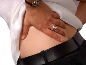
Osteochondrosis of the lumbar spine is indicated by pain in the lower back. Due to staying in an uncomfortable position for a long time, the pain may increase. After sleeping or resting on your back, the pain decreases or disappears completely.
The occurrence of stressful conditions for the body can cause the development of an acute condition. Often this happens during hypothermia, heavy loads, and sudden movements. Exacerbation is characterized by severe pain, which from the lumbar region can go down to the legs. The body can freely try to reduce the load on the affected part of the spine with strong tension in the lower back muscles. A patient with osteochondrosis of the lumbar spine tries to find a comfortable position where the pain decreases.
Diagnosis of lumbar osteochondrosis
Diagnosis of osteochondrosis is carried out in several stages. The doctor needs to talk to the patient, ask about the complaint, find out about the nature of the pain, where it is localized, at what time painful sensations make themselves felt, their duration, intensity, etc. In addition, the doctor will know under what conditions the pain appears, when it gets stronger or less.
After this, the vertebrologist examines the anamnesis, i. e. disease history. The doctor will definitely explain how long the painful condition lasts, what is the cause of the pain, and how you feel during the period of discomfort. The important thing is early treatment, as well as the effectiveness of certain treatment methods. A qualified doctor will certainly ask the patient about living and working conditions, range of motion, well-being under certain loads, and past illnesses. It is very important to find out if the patient has an injury to the spine, if he plays sports, and if any close relatives have spinal diseases.
The next step in the diagnosis is the examination of the patient. The doctor will pay attention to the position of the head, legs and arms in relation to the body, the gait, the way of holding, the symmetry of the body area, the condition of the skin in the damaged area, and the movement of the patient. After this, the range of motion of the spine and the degree of damage are established. To do this, the doctor will ask the patient to bend forward, sideways, backwards, ask him to move different parts of his back, and tilt his head. Healthy people should not experience any irritation or pain in their joints during the simple test.
If the patient does not have osteochondrosis, then he can reach his chest and shoulders with his chin. Head movement in each direction is about 60 degrees. A 45-degree angle is formed by bending sideways between the head and the upper cervical spine. The distance from the sacrum to the spinous process of the seventh cervical vertebra increases by 5-7 centimeters when bending forward. This distance is reduced by 5-6 cm when bending backwards. An experienced vertebrologist will pay attention to how the knee and hip joints participate in bending, and how the configuration of the spine changes.
Treatment of osteochondrosis of the lumbar spine
Lumbar osteochondrosis requires complex, intensive and long-term treatment. This is especially true for chronic cases with multiple intervertebral hernias and protrusions.
Effectivetreatment of osteochondrosis of the lumbar spineachieved with a reflex method that does not cause side effects, but brings maximum benefits. Remember, recovery from such a serious illness cannot come quickly. In each case of lumbar osteochondrosis, the doctor prescribes an individual treatment regimen.
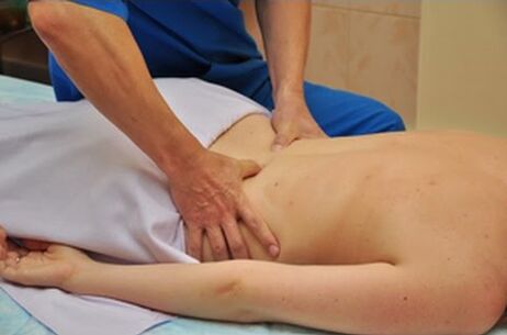
Keep in mind that manual therapy can only be used in cases where the patient's spine is not damaged by protrusions and intervertebral hernias. The integrated use of this technique allows you to restore normal blood microcirculation, relieve congestion, vascular edema, relieve muscle spasms, restore the balance of metabolic processes in the vertebral tissue and intervertebral disc, and also improve tissue nutrition in the lumbar region. As a result, the natural regeneration process is stimulated. It should be remembered that in the case of lumbar osteochondrosis, manual therapy should be aimed at improving the function of the spine.
Treatment procedures for lumbar osteochondrosis are complemented by the use of herbal medicines that increase conservation in the body and also restore the balance of metabolic processes. The vertebrologist will recommend dietary correction and a more active lifestyle to the patient. It is important to adjust your weight, because excess weight puts additional pressure on the lower back and also worsens the development of osteochondrosis of the lumbar spine.
Experienced specialists who treat osteochondrosis allow patients to achieve serious results, as well as avoid surgery, improve motor activity, eliminate lower back pain, and comprehensively improve the body to prevent further deterioration. Acute pain disappears after 1-3 sessions of manual therapy, and the positive effect of treatment is achieved on average after 10-15 sessions. Remember that the start of treatment on time is a guarantee to achieve a positive result.
During the period of exacerbation of pain in the lower back, the patient is recommended to stay in bed for 2-3 days. To quickly relieve pain, nonsteroidal anti-inflammatory drugs, pain relievers, venotonics, diuretics, nicotinic acid, and B vitamins are prescribed. Walking with crutches, dry or underwater traction on the spine can also be prescribed to unload the spine. If necessary, blockade with glucocorticoids or paravertebral blockade with anesthetic is prescribed.
Physiotherapy treatment of osteochondrosis of the lumbar spine may also include electrophoresis, laser therapy, and ultraviolet radiation. Physical therapy is a mandatory component of a comprehensive treatment program. Some exercises can be used in the acute stage, but almost all exercises are recommended to be done after the pain subsides. Thanks to regular physical exercise, tissue nutrition is restored, blood and lymph supply to the intervertebral disc improves, and the flexibility and elasticity of joints and cartilage is gradually restored. Thus, the patient's condition improves, and the interval between periods of exacerbation lengthens.
Spa treatments also have a positive effect on health. In climatic resorts, iodine-bromine, hydrogen sulfide and radon baths are prescribed.
Surgical treatment methods are used only if the patient's pain cannot be eliminated by conservative methods for a long time, with paresis of the muscles of the lower leg and disruption of the natural process of urination and defecation. During the operation, the herniated disc is removed and the spinal segment is strengthened.
Prevention of lumbar osteochondrosis
Prevention of lumbar osteochondrosis consists of following simple rules recommended by vertebrologists. Remember that the development of the disease can only be prevented by carefully following these rules, no matter what:
- Keep your lower back dry and warm, don't overcool your spine, and avoid drafts.
- Do not lift heavy objects or carry them over long distances.
- Try not to make sudden movements.
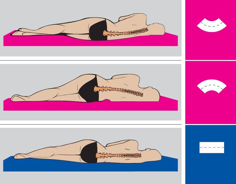
- Maintain proper posture while working and resting.
- Change your position as often as possible, try not to stay in one position for a long time.
- Do physical therapy.
- Try not to stay in a bent position for a long time.
- When cleaning, use long mops, brooms, and vacuum cleaners with long tubes so you don't have to bend over.
- Lift the load correctly: bend with a straight back, or bend your knees, lift the bag with a straight back and stand up straight. Keep your hands with the weight as close to your body as possible.
- If you need to bend low to pick something up off the floor, such as under a table or bed, get down on one knee and keep your back straight.
- Distribute the weight evenly between the two hands.
- Strengthen your gluteal muscles, stretch your spine, take a walk every day.
- Balance your diet, enrich your diet with dairy products and plants.
- Stick to the drinking regime - 1. 5-2 liters of water and herbal tea every day.
- Get rid of bad habits - alcohol, smoking, drugs.